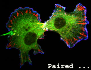
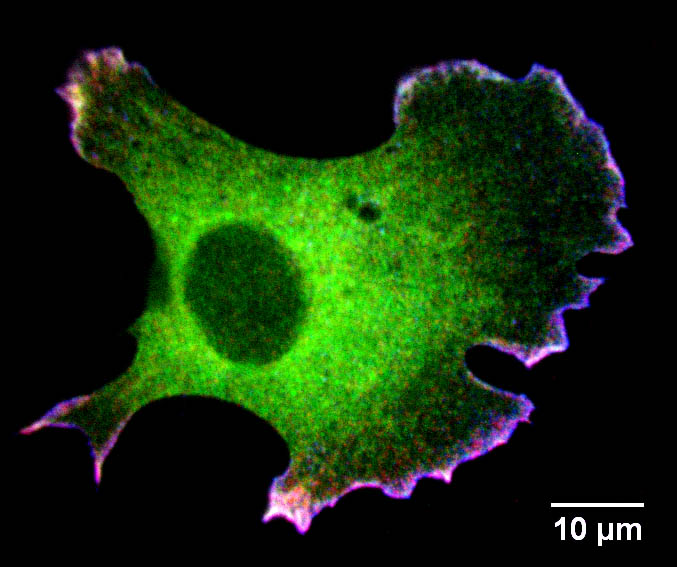
 The origin of these images
In Bear laboratory, we were studying actin based cellular events. For me, I was focusing on fibroblast lamellipodia dynamics. One of the basic approaches I was using is immunofluorescent staining and spinning disk confocal imaging. I have collected many very pretty images, which I would like to share with you :-)
 Usually, I immunostained cells following Bear Lab's protocol. Then use spinning disk confocal microscope to take z-stack images with 0.2 µm interval. After that, I use ImageJ's "extended depth of focus" plugin (not "extended depth of field") to combine information from each frame to generate in-focused images, which does much better job than the traditional maximal intensity projection. Here are examples of fixed-and-stained fibroblasts.
You can follow this link and find more "pretty" images.